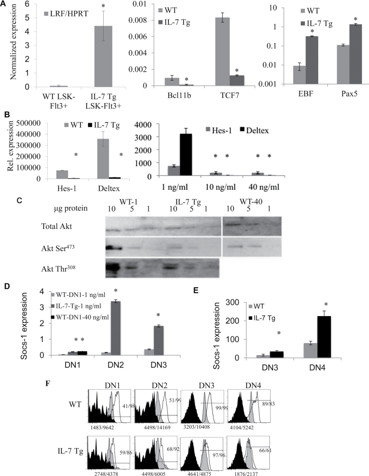
Fig. 3.
Gene expression analysis of cells exposed to high IL-7 doses. (A) Panels compare the relative gene expression of IL-7 Tg and WT. Left panel, LRF expression in LSK-Flt3+ cells; middle panel, Bcl11b and TCF-7 expression in DN1 c-Kit+ cells; right panel, EBF and Pax5 expression in DN1 c-Kit+ cells (*P < 0.001). (B) Left panel, relative expression levels of Hes-1 and Deltex expression in DN1 c-Kit+ cells from WT and IL-7 Tg mice (*P < 0.0001 for Hes-1 and 0.0008 for Deltex). Right panel, relative expression levels of Hes-1 and Deltex expression in DN1 c-Kit+ cells sorted from WT LSK-Flt3+ and cultured with OP9-DL1 cells at 1, 10 or 40ng ml−1 of IL-7 (Hes-1 *P < 0.05, Deltex *P < 0.0001). Data shown in (A) n = 8–9 mice each group done in triplicate and (B) are from two experiments done in triplicate. (C) LSK-Flt-3+ progenitors were sorted from IL-7 Tg and WT mice. WT cells were cultured on OP9-DL1 stromal cells with 1ng ml−1 (WT-1) or 40ng ml−1 of IL-7 (WT-40). IL-7 Tg cells were cultured on OP9-DL1 stromal cells with 1ng ml−1 IL-7 (IL-7 Tg). After culture, DN1 c-Kit+, DN2 c-Kit+ and DN3 c-Kit− progenitors were separately sorted then mixed in the same proportions for each group. Total and phospho-Akt Ser473 are shown for all groups. Phospho-Akt-Thr308 is shown in IL-7 Tg and WT-derived cells. (D) Normalized SOCS-1 expression in sorted DN1-c-Kit+, DN2-ckit+ and DN3-c-Kit− from WT LSK-Flt-3+ and OP9-DL1 cell co-cultures treated with1ng ml−1 or 40ng ml−1 of IL-7. IL-7 Tg LSK-Flt3+ were cultured with 1ng ml−1 of IL-7 (*P < 0.0001). (E) Normalized SOCS-1 expression in sorted DN3-c-Kit− and DN4-c-Kit− thymocytes from IL-7 Tg and WT mice (*P = 0.0004; **P = 0.0001). (F) STAT5 intracellular staining in DN thymocytes from IL-7 Tg and WT mice. The black filled histograms show the isotype control for STAT5 staining; the grey histograms show STAT5 staining for unstimulated cells; the open histograms correspond to STAT5 staining in progenitors stimulated with IL-7. Gates were DN1-c-Kit+, DN2-ckit+, DN3-c-Kit− and DN4-c-Kit− within the Lin− population. The experiment was repeated three times.

