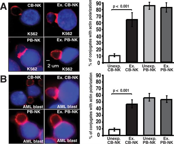
FIGURE 3.
Freshly isolated unexpanded CB-NK cells exhibit decreased immunological synapse formation that can be reversed with ex vivo IL-2 expansion. A, Left panel, confocal fluorescence microscopic images show representative examples of unexpanded and expanded (Ex.) CB- and PB-NK conjugates with K562 cells (stained blue with CMAC-dye). Images show one medial optical section in the z-plane. Original magnification 40×. The right panel shows percent of conjugates with F-actin polarization (red) corresponding to the synapse images shown on the left. B, Left panel, confocal fluorescence microscopic images of unexpanded and expanded (Ex.) CB- and PB-NK conjugates with AML patient primary blasts (blue). The right panel shows the percentage of F-actin accumulation at the NK immune synapse. Values are the means ± SEM from six independent experiments, with at least 50 random conjugates analyzed per experiment.

