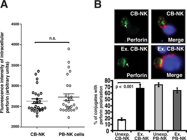
FIGURE 6.
Ex vivo IL-2 expansion of CB-NK cells promotes polarization of perforin to the lytic mature synapse. A, Quantitative analysis of intracellular perforin expression levels in non-conjugated CB- and PB-NK cells using confocal microscopy and image analysis is shown. Fluorescence intensity was calculated using ImageJ software. Data are representative of at least six independent experiments. Statistical analyses were performed using the nonparametric Mann-Whitney test using PRISM software. B, Unexp. and Ex. CB- and PB-NK conjugates with target AML blasts (stained blue) were scored for polarization of perforin (stained green) at the NK cell immune synapse (F-actin stained red). The upper panel shows representative confocal fluorescence microscopic images of unexpanded (Unexp.) and expanded (Ex.) CB conjugates with AML blasts. Arrows indicate protein localization at the NK cell-AML blast target cell synapse site. Co-localization of proteins in the merged images is shown in yellow. Original magnification 40×. The lower panel shows the percent of conjugates with perforin polarization of unexpanded and expanded CB-NK cells (corresponding to the synapse images shown in the upper panel) as well as unexpanded and expanded PB-NK cells. Values are the means ± SEM from six independent experiments, with at least 50 random conjugates analyzed per experiment.

