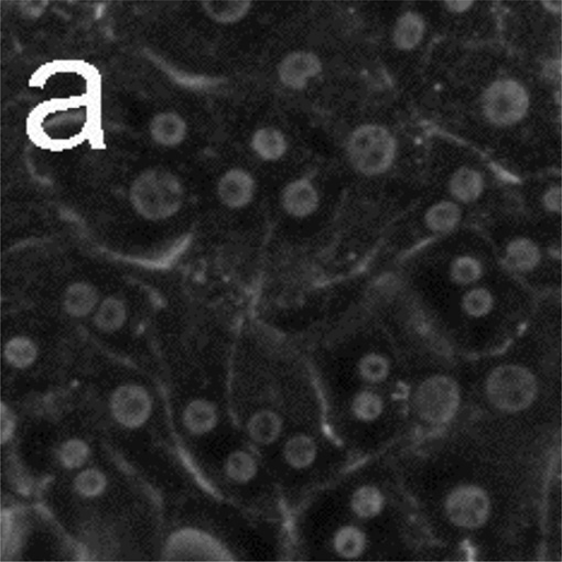
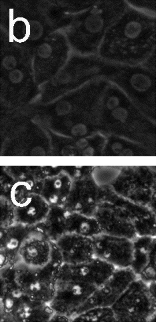
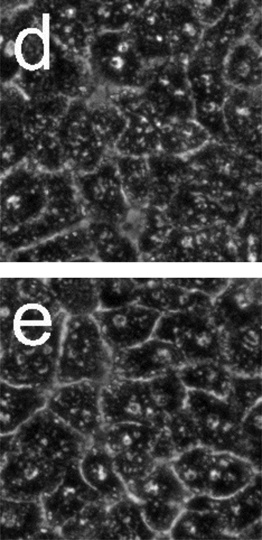
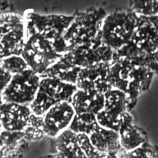
Figure 3.
Phase-contrast microscope images of primary rat hepatocytes seeded on tissue-culture polystyrene multi-well plates. (a) Hepatocytes without toxin at t = 0. (b) Hepatocytes without toxin after 18 h. Hepatocytes after exposure to 50 µM Cd2+ at (c) 2 h, (d) 6 h, (e) 10 h, and (f) 14 h. Time 0 corresponds to the time in which toxin (or a blank for the control) was added. Cells exposed to Cd2+ begin to show granular appearance within the first 2 h. Most cellular features become indistinguishable 14 h after Cd2+ exposure. Image dimensions are 150 µm × 150 µm.


