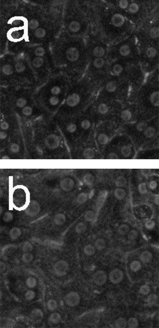
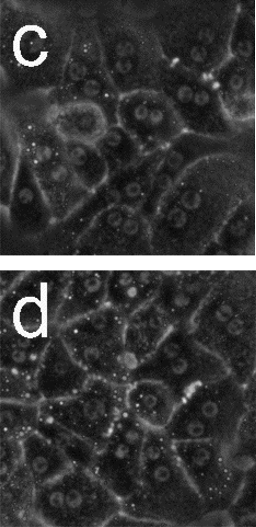
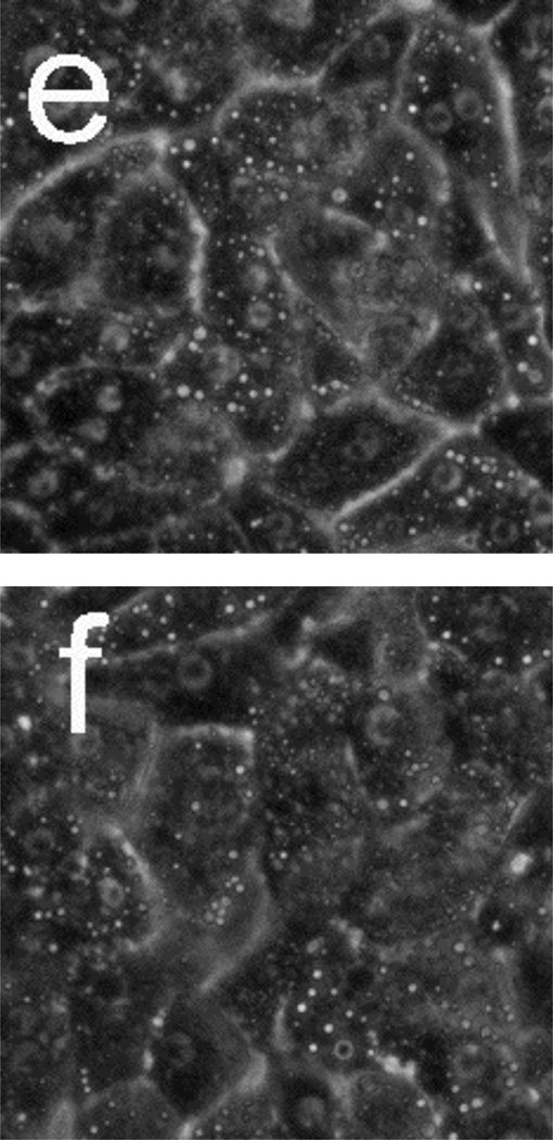
Figure 5.
Phase-contrast microscope images of primary rat hepatocytes seeded on tissue-culture polystyrene multi-well plates after exposure to 10 mM N-acetyl-p-aminophenol (APAP) for (a) 2 h and (b) 18 h, and 40 mM APAP for (c) 4 h, (d) 8 h, (e) 12.5 h, and (f) 18 h. There is little to no change in cellular morphology after exposure to 10 mM APAP for 18 h. However, 4 h after exposure to 40 mM APAP, the presence of lipid droplets (circular, phase-bright features) becomes evident. The number of lipid droplets continues to increase up to 12.5 h. There is significant granular appearance 18 h after introduction of 40 mM APAP, although some cellular features are still apparent. Image dimensions are 150 µm × 150 µm.


