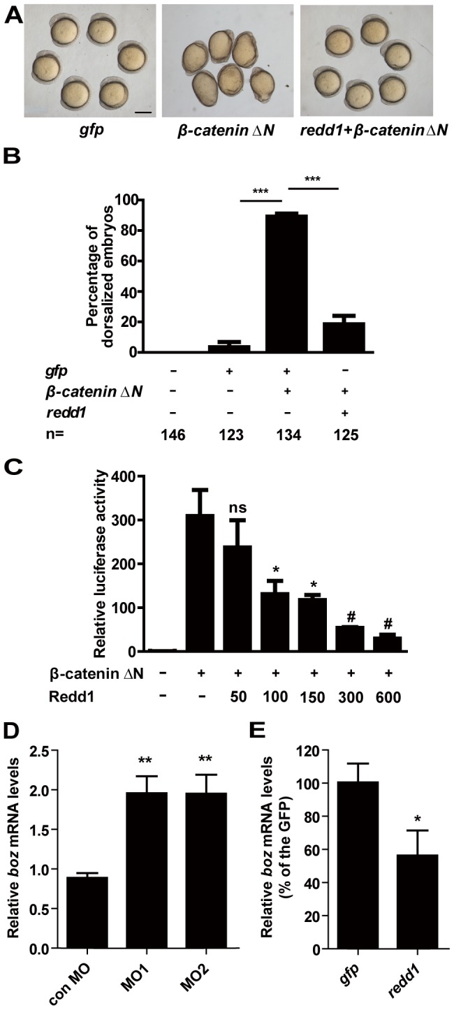
Figure 5. Redd1 inhibits β-catenin action.

A and B) Redd1 inhibits β-catenin action in vivo. Representative view of gfp mRNA-, β-catenin ΔN mRNA-, and β-catenin ΔN mRNA + redd1 mRNA-injected embryos at 5 somite stage is shown in A). Scale bar = 200 µm. Quantitative results are shown in B). The results are from three independent experiments and the total embryo number is given at the bottom. *** P<0.001. C) Redd1 inhibits β-catenin activity in vitro. HEK293T cells were transfected with β-catenin ΔN plasmid DNA and increasing doses of Redd1 plasmid DNA, together with the same amount of TCF/LEF-luciferase reporter DNA. Cells transfected with TCF/LEF-luciferase reporter DNA alone were used as negative control (−).Values are means ± S.E., n = 3. ns, not significant, * and #, P<0.05 and P<0.001 compared to the β-catenin ΔN group. D) Redd1 knockdown increases boz expression. Embryos were injected with control MO, redd1 targeting MO1 or MO2 at one-cell stage. The embryos were raised to the dome stage. The boz mRNA levels were measured by quantitative real-time RT-PCR. E) Forced expression of Redd1 decreases boz expression. Embryos were injected with gfp mRNA or redd1 mRNA at one-cell stage and were analyzed at dome stage. The boz mRNA levels were measured by quantitative real-time RT-PCR.
