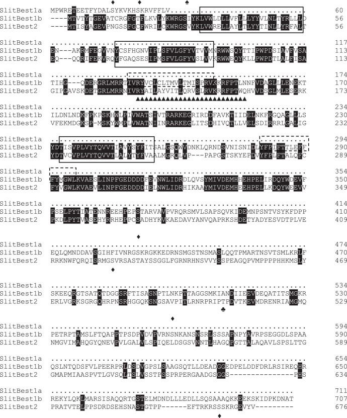
Figure 1. Alignment of S. littoralis bestrophins proteins.
Alignment was realized with ClustalW2 (http://www.ebi.ac.uk/Tools/clustalw2/) and displayed with BoxShade (http://mobyle.pasteur.fr/cgi-bin/portal.py?) The amino acid sequence of SlitBest1a that matches with SlitBest1b is omitted. Identical amino acids between all sequences are marked in black. Predicted transmembrane domains are surrounded by a solid line for domains shared by the three sequences and by a dotted line for the Slitbest1a and SlitBest1b transmembrane domains. Predicted PKC, PKA and PKB phosphorylation sites are indicated by diamonds (♦), spades (♠) and clubs (♣), respectively. The bestrophin RFP domain is indicated by triangle (▴).

