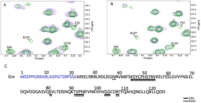
Figure 5. Fragments of 15N-1H HSQC spectra of 15N-labeled reduced Grx domain of mouse TGR titrated with unlabelled GSH and GSSG (panels A and B, respectively).
Green corresponds to free Grx and magenta to Grx incubated with GSH/GSSG. Only the residues for which alteration of NMR parameters upon titration was observed are marked. Panel C: qualitative representation of the data. Solid and dashed horizontal lines below the Grx amino acid sequence highlight the residues interacting with GSH and GSSG, respectively.The N-term of Grx is marked in blue. For more details see the text.

