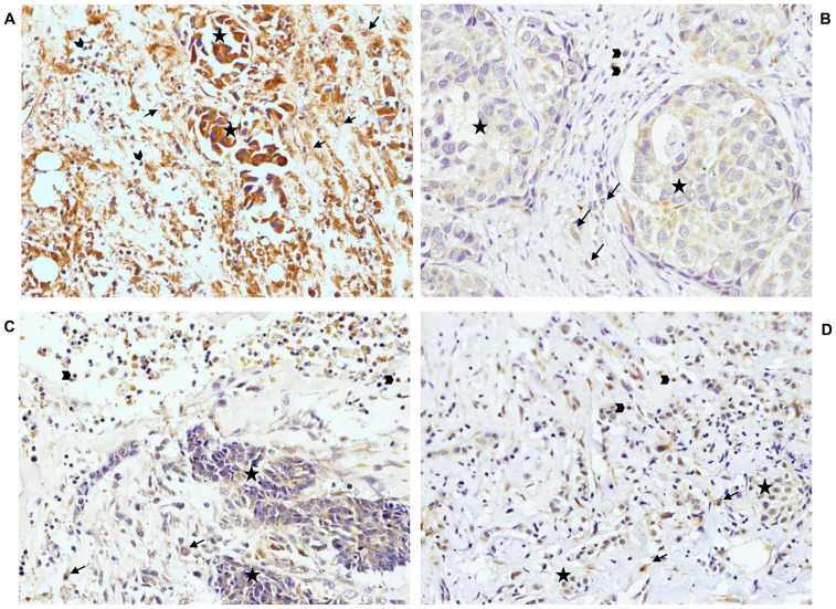
Figure 3. Representative example of immunostaining.
MMP11 (A) and TIMP2 (B) immunostaining at the tumor center and MMP9 (C) and MMP14 (D) at the invasive front (×200 magnification), indicating the different cell types. Tumor cells (★), lymphocytes (<$>\raster(70%)="rg2"<$>) and macrophages (<$>\raster(70%)="rg1"<$>).

