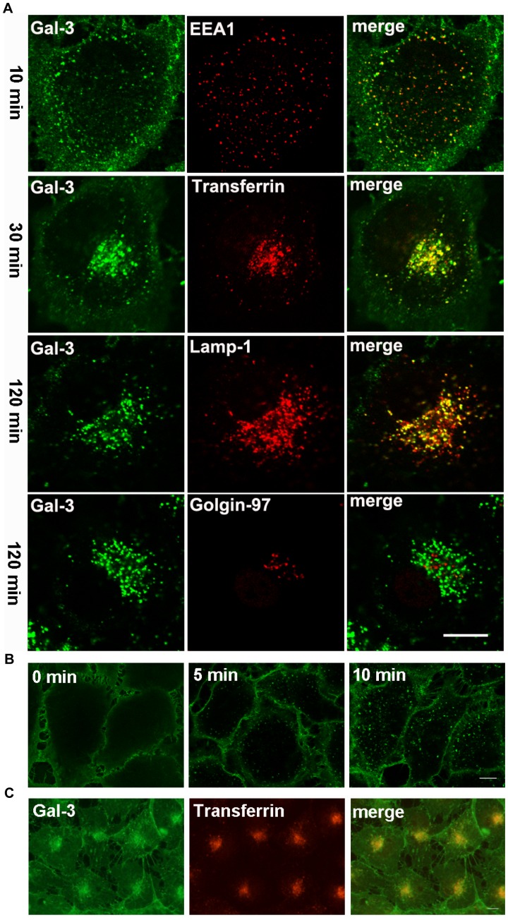
Figure 4. The endocytic pathways of Gal-3 in HUVEC.
A: The cells were incubated with DTAF-Gal-3 and Cy3-transferrin (only at 30 min) at 37°C for 10, 30, or 120 min and were then processed for IF analysis with antibodies against EEA1, Lamp-1 and Golgin-97. B: The cells were incubated with DTAF-Gal-3 at 4°C for 60 min. After washing, the cells were either fixed immediately (0 min) or transferred to 37°C for 5 min or 10 min and then fixed. C: The cells were incubated with DTAF-Gal-3 and Cy3-transferrin at 20°C for 60 min. The images were obtained with a confocal microscope (A and B) or a fluorescence microscope (C). Scale bar, 10 µm.

