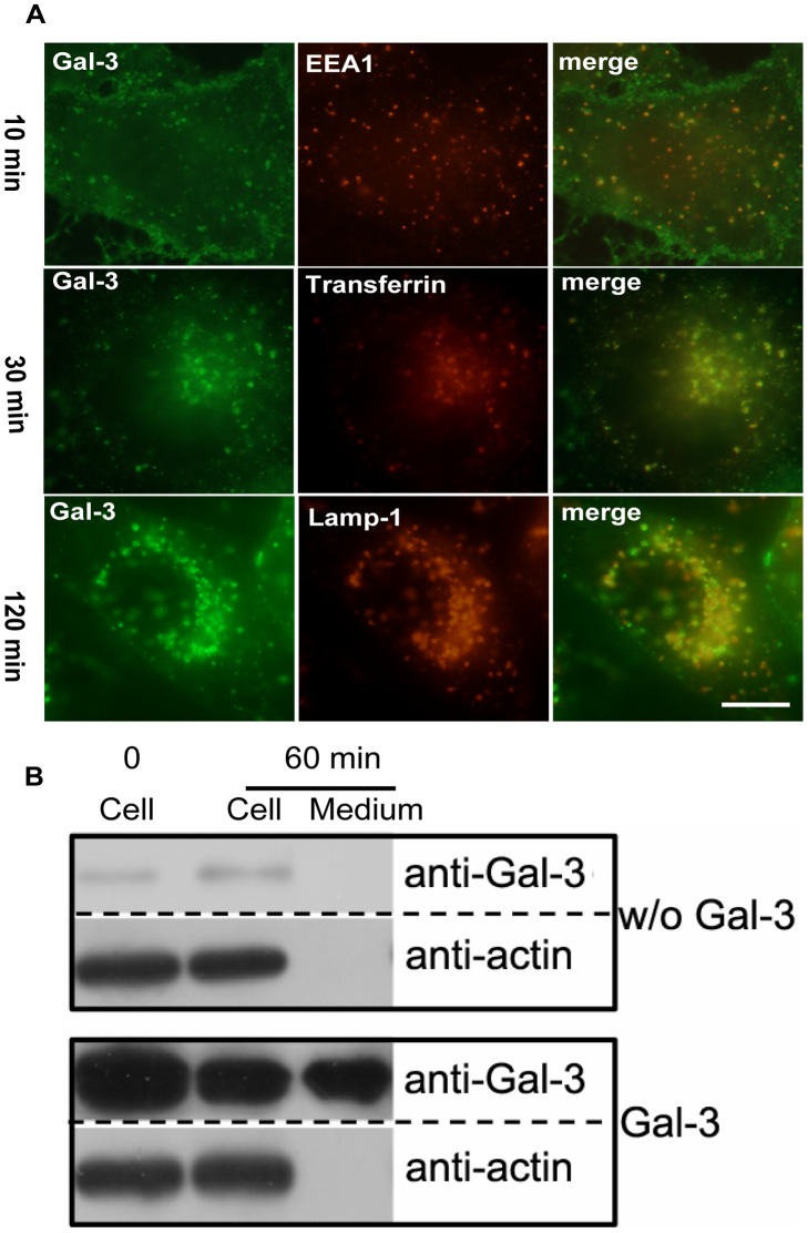
Figure 9. The two endocytic pathways of Gal-3 observed in HUVECs are also in HMEC-1 cells.
A: Fluorescent images showing the kinetics of Gal-3 endocytosis. DTAF-Gal-3 was incubated with HMEC-1 for 10 min, 30 min, or 120 min at 37°C and then processed for IF analysis as described in Figure 4. The images in this figure were obtained with a fluorescence microscope. Scale bar, 10 µm. B:Western blot showing the exocytosis of Gal-3. The HMEC-1 cells were incubated with or without (w/o) Gal-3 at 4°C for 30 min. After extensive washing, the cells were either immediately lysed (0 min) or incubated in fresh SFM at 37°C for 60 min. After incubation, both the medium and the cells were analyzed with western blotting.

