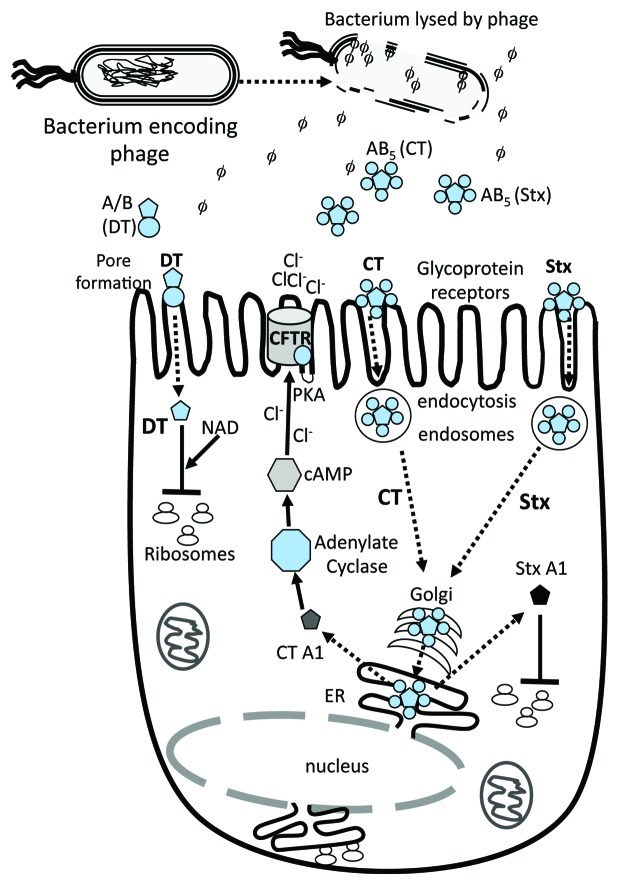
Figure 1. Schematic of toxins function in eukaryotic cells. Dotted arrows indicate steps in bacterial lysis with phage/toxin release, toxin uptake by host cell and target activity. CTXphi which encodes cholera toxin (CT) does not lyse its bacterial host cell. Black solid arrows or t-bars represent activation or blocking of specified bacterial function by toxin. AB denotes a two subunit protein, subunit A is the catalytic subunit and B is the receptor binding or docking subunit on target cell. AB5 denotes a toxin that contains one A subunit and 5 B subunits. Three examples are given; the A/B diphtheria toxin (DT) and the AB5 toxins CT and shiga toxin (Stx). DT gains direct entry into target cell by pore formation. The second method of toxin uptake is by receptor mediated endocytosis, a mechanism used by CT and Stx. Only the A subunit of DT enters the cell cytosol and along with NAD, ADP-ribosylates Elongation factor 2 blocking protein synthesis. Both CT and Stx are retrograde trafficked to the Golgi apparatus. In CT, the A subunit is dissociated from B subunits in the endoplasmic recticulum (ER) and released into the cytosol where it activates adenylate cyclase increasing cAMP which in turn activates PKA resulting in the CFTR membrane Cl- channel activation. The A subunit of Stx ADP- ribosylates the 28S rRNA ribosomal subunit, thus blocking protein synthesis.

An official website of the United States government
Here's how you know
Official websites use .gov
A
.gov website belongs to an official
government organization in the United States.
Secure .gov websites use HTTPS
A lock (
) or https:// means you've safely
connected to the .gov website. Share sensitive
information only on official, secure websites.
