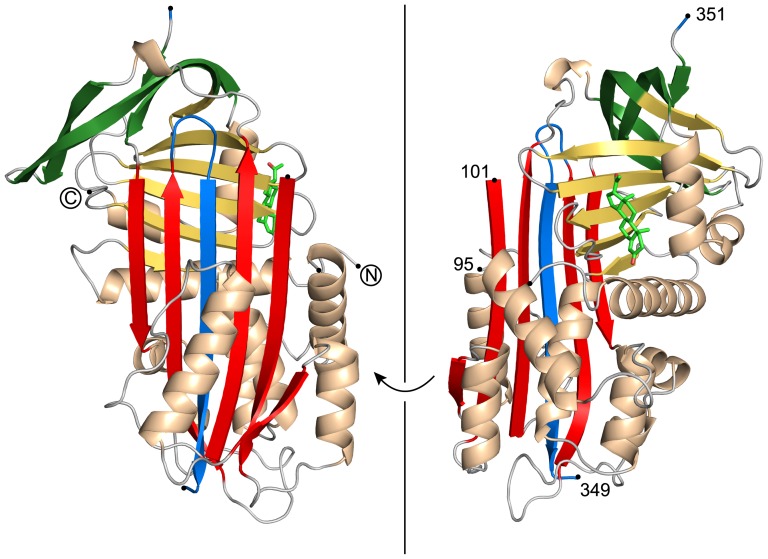
Figure 1. Crystal structure of cleaved human CBG in complex with progesterone.
The central β-sheet A is shown in red, with the inserted RCL as part of it in blue. β-sheet C and β-sheet B are colored in green and yellow, respectively. On top of β-sheet B the bound steroid hormone progesterone is depicted in bright green. Electron density for residues 96 to 100 and residue 350 is missing. Chain breaks are shown with black dots.

