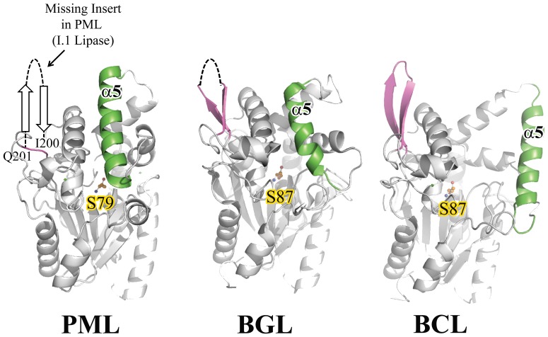
Figure 2. Comparison of the distinguishing β-turn-β structure that differentiates family I.1 lipases from family I.2.
The position of the absent insertion in PML is shown. The ∼14 residue insertion in BGL and BCL (family I.2 lipases) is colored purple. As reference, lid helix α5and the catalytic serine are shown and colored green and orange respectively.

