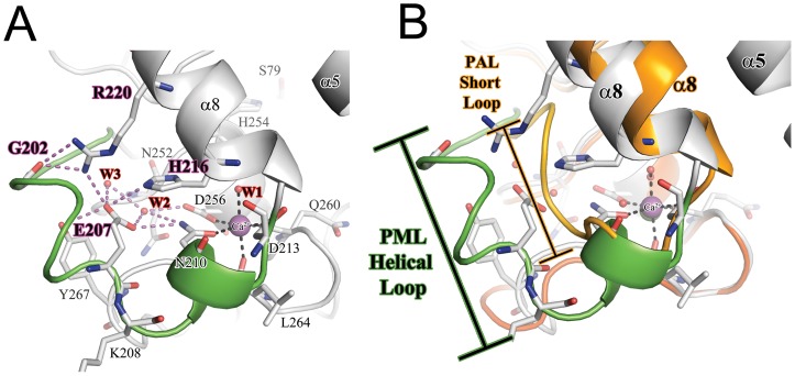
Figure 3. The structure of Ca2+ binding site and supporting loop region in PML.
A) A detailed view of the C2+ binding site. The Ca2+ ion is shown as a purple sphere. The coordination of residues to Ca2+ are shown as black dashes. The intricate H-bonding network connecting the loop to lid helix α8 are shown as purple dashes. The helical loop region unique to P. mirabilis and other Proteus/psychrophilic subfamily I.1 lipases is shown in green. B) Comparison of the loop region in P. mirabilis (PML; green) and P. aeruginosa (PAL) orange. The loop that precedes the Ca2+ binding site is 5 residues longer in PML compared to PAL.

