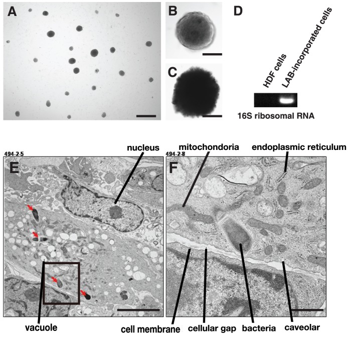
Figure 1. Characterization of LAB-incorporated cell clusters.
(A) Typical LAB-incorporated cell clusters occurring on adherent culture dishes. (B) Characteristics of LAB-incorporated cell clusters. (C) ALP staining of LAB-incorporated cell clusters. (D) RT-PCR on LAB-incorporated cell clusters after 12 days of incorporation using a 16S ribosomal RNA primer set. (E) Ultrastructural picture of LAB-incorporated cells. (F) Hyper-magnification of a square in (E). Scale bars: 1 mm in (A), 100 µm in (B) and (C), 5 µm in (E), and 1 µm in (F).

