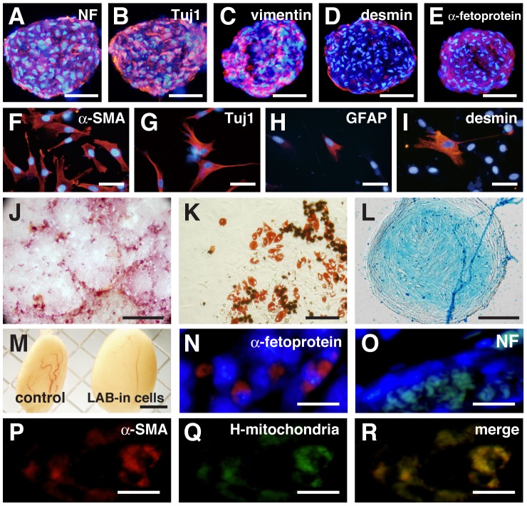
Figure 3. Multiple-lineage differentiation of LAB-incorporated cell clusters.
(A, B) The floating culture for the neural induction produced clusters that expressed the neural markers neurofilament (NF, A) and Tuj1 (B). (C–I) Differentiating LAB-incorporated cell clusters cultured in the floating condition expressed vimentin (C), desmin (D), and α-fetoprotein (E). Differentiating LAB-incorporated cell clusters cultured in the attached condition expressed α-SMA (F), Tuj1 (G), GFAP (H), and desmin (I). (J) Induced osteocytes were stained with Alizarin Red S. (K) Induced adipocytes were stained with Oil Red O. (L) Induced chondrocytes were stained with Alcian Blue. (M) Uninjected testis and testis injected with LAB-incorporated cell clusters (12 weeks). (N, O) Immunocytochemistry of α-fetoprotein (N) and NF (O) in testis injected with LAB-incorporated cell clusters. (P–R) Double-staining of α-SMA (P) and human mitochondria (Q). Scale bars: 100 µm in (A–E), 50 µm in (F–I), 500 µm in (J), 100 µm in (K), 50 µm in (L), 200 µm in (M), and 20 µm in (N–R).

