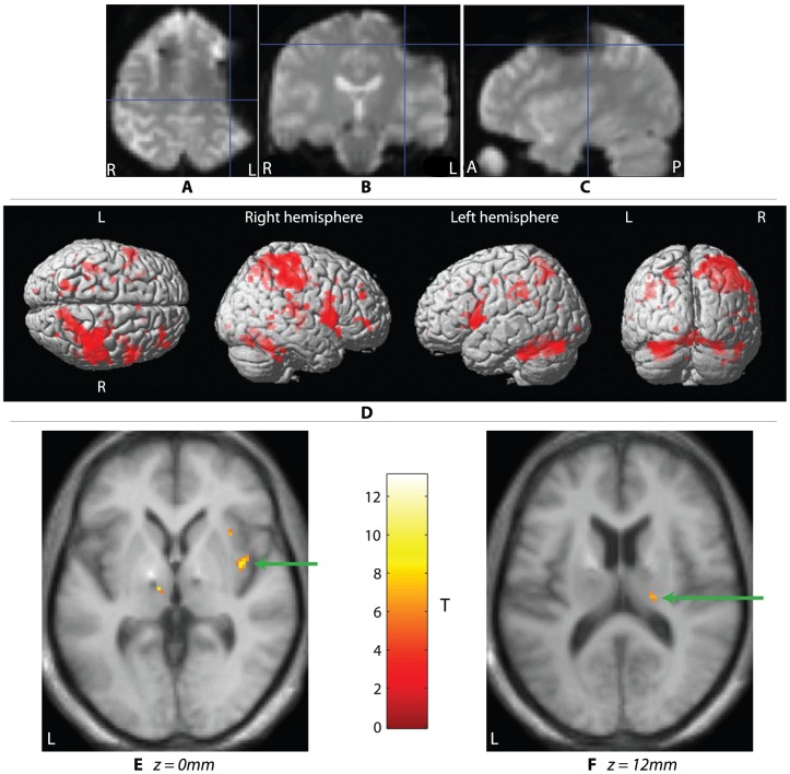
Figure 2. Imaging results.
A typical drop-out artefact in a single subject's GE-EPI acquisition viewed from (a) axial, (b) coronal, and (c) sagittal sections; cross-hair position = −34.8, −21.5, 53.3 mm (MNI coordinates). SPMs in (d) summarize the movement network on a rendered MNI brain (p<0.001 uncorrected). Clusters representing BOLD signal increases in the insula cortex (e, green arrow), and thalamus (f, green arrow).

