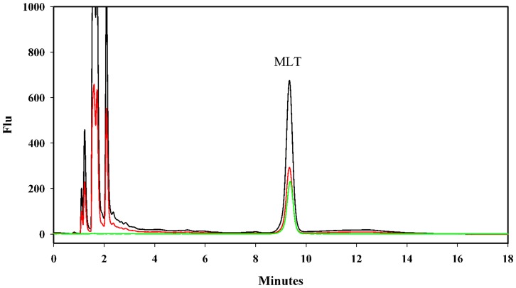
Figure 1. The detection of melatonin by HPLC.
Overlaid chromatograms of a melatonin standard (green, 1×10⁻⁸ gr mL−1) and a sea anemone tissue extract (N. vectensis) with (black) and without (red) melatonin enrichment (1×10⁻⁸ gr mL−1). Chromatographic separation of tissue extracts was conducted based on fluorimetric detection (λex = 280 nm and λem = 345 nm). The mobile phase consisted of a mixture of 0.1% (v/v) formic acid in acetonitrile:water 17∶81 (v/v), which was delivered isocratically at a flow-rate of 1 mL min−1. The detailed conditions are described in the Materials and Methods.

