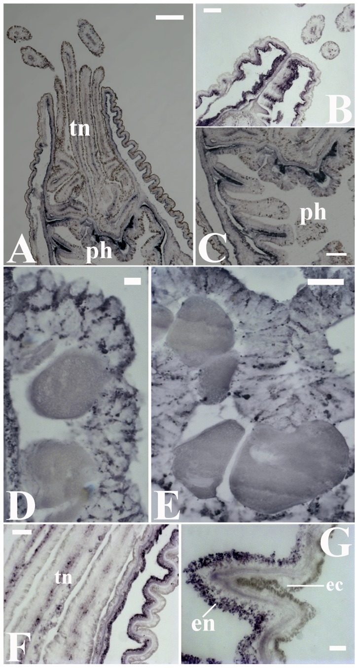
Figure 5. The expression pattern of hydroxyindole-O-methyltransferase (HIOMT) mRNA in Nematostella vectensis.
Two representative HIOMT orthologs were evaluated using in situ hybridization (ISH) with specific probes. (A–C) The HIOMT expression pattern was similar between orthologs and indicated that melatonin is predominantly produced in the circumference of the actinopharynx (ph). High HIOMT expression levels were also evident in the endodermal layer of the hypostome (retracted individual, B). A higher magnification image of the outer surface of the endothelium in the actinopharynx folds (C). (D, E) Substantial HIOMT expression in reproductive tissues suggested that the considerable level of the melatonin that is observed in these tissues (see Fig. 1E–F) is locally produced. A uniform HIOMT expression pattern was observed among the cells. (F, G) In the body wall, predominantly endodermal HIOMT expression was observed throughout the apical end of the anemone and was uniformly distributed throughout the cells. This pattern differed from the pattern of melatonin immunoreactivity (see Fig. 1B–C). Note that melatonin production occurs also in tentacles (tn), albeit at lower levels. Endoderm (en), ectoderm (ec). Scale bars: A = 200 µm; B, C = 100 µm; E, F = 50 µm; D, G = 20 µm.

