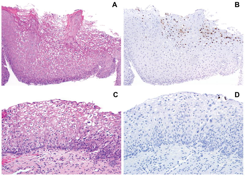
Figure 1.
A, C. Low grade squamous intraepithelial lesion (LSIL/CIN 1). B. Extensive L2 expression in the superficial epithelial layers; L1 showed similar findings (not shown). D. Limited L1 expression (two positive nuclei) in the most superficial epithelial layer; the distribution of L2 expression was identical (not shown).

