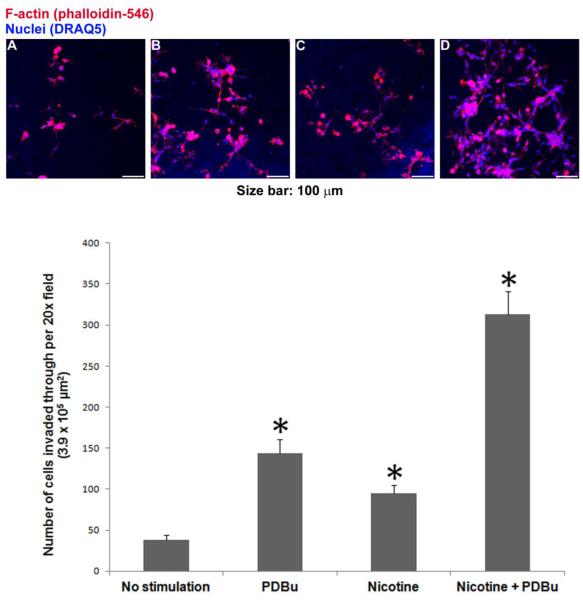
Figure 8.
Nicotine and PDBu-induced invasion of human aortic smooth muscle cells through extracellular matrix, as measured by matrigel-coated transwell invasion assay. Top panels A to D show representative images of cells reaching the bottom side of the membrane under the following four experimental conditions: (A) No Stimulation: neither nicotine nor PDBu was added to the top or bottom chamber; (B) PDBu: 2 μM PDBu in the bottom chamber only; (C) Nicotine: 2 μM nicotine in both top and bottom chambers; and (D) Nicotine + PDBu: 2 μM nicotine in top chamber; 2 μM nicotine and 2 μM PDBu in the bottom chamber. Fluorescence from DRAQ5-labeled nuclei appeared as blue, whereas Alexa Fluor 546-conjugated phalloidin-labeled filamentous actin appeared as red. For each membrane, the number of cells in the 20x microscopic field was counted based on DRAQ5-labeled nuclei. Bottom graph shows cell counts in mean + one standard deviation (n = 3). Asterisks indicate significant difference from control (no stimulation). In addition, two-way ANOVA was performed to analyze individual and interactive effects of PDBu and nicotine on cell invasion.

