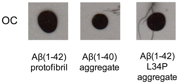
Figure 2.
OC-negative monomer fractions develop OC immunoreactivity. Aβ(1-40) and Aβ(1-42) L34P monomer fractions from Figure 1 were incubated quiescently at 37° C for 6 and 13 days respectively and subjected to dot blot analysis. For comparison, an Aβ(1-42) protofibril fraction eluted in the Superdex 75 void volume in F-12 medium without phenol red was also analyzed. 2 μl of each sample at final concentrations of Aβ(1-42) (28 μM), Aβ(1-40) (39 μM), and Aβ(1-42) L34P (20 μM) was used.

