
Figure 1.
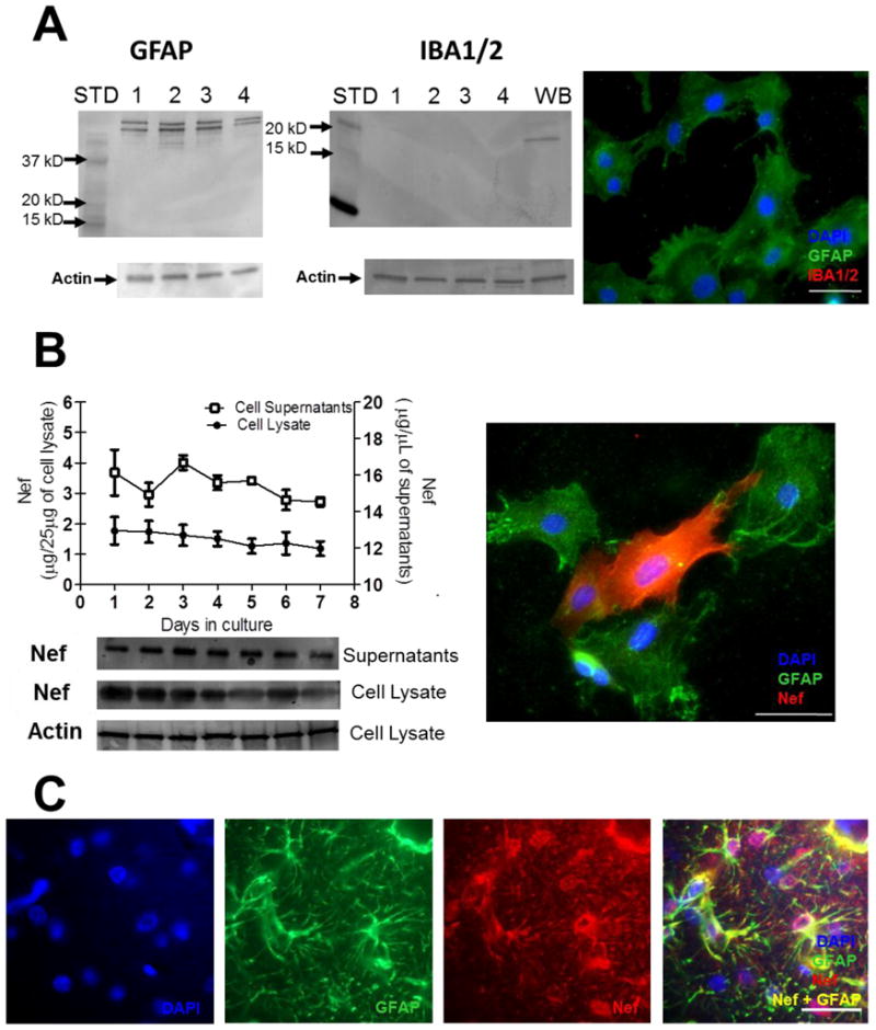
Nef is expressed for seven days after transfection of primary rat astrocytes in vitro and in vivo from infused cells. (A) Primary culture composition was assessed for astrocytes (GFAP) and microglia (Iba1/2). Western blot of three, independent primary cell extractions (lanes 1–3) and SVGA astrocytes (lane 4) show positive for GFAP and negative for Iba1/2. Positive control for microglia in whole brain (WB) tissue shows positive reaction with Iba1/2. Molecular weight markers are labeled (STD). In the third panel, immunofluorescence for GFAP (green) and Iba1/2 (red) show cultures are astrocytes. DAPI was used for nuclear counterstaining. (B) Cells and culture supernatants were collected each day for seven days after transfection with Nef plasmid. Western blots, lower part, measured Nef protein expression. Densitometry of western blots was used to quantify (upper part) Nef from cell lysates and supernatants from the same cell cultures. Concentrations of Nef were estimated using a Nef protein standard (refer to methods). Actin served as a loading control for cell lysates. Immunofluorescence indicates Nef (red) expression from GFAP positive (green) astrocytes. (C) Immunostaining of hippocampal tissue proximal to infusion site: left to right, DAPI, GFAP (green), and Nef (red) in split channel, and merged image at seven days post-surgery. Scale bars = 25μM.