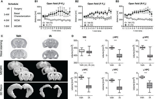
Figure 4.
Consequences of ibotenic acid lesions. (A) Schedule after surgery [d (day), week (W); for a summary of the results of the basal characterization see Table 1]. Panel (B1–B3) There were three independent batches of sham-operated (Veh) or ibotenic acid-lesioned (IA) mice. Veh controls habituated to the open field or at least stayed at a constant exploration level, whereas IA mice even showed a sensitized response in a highly replicable manner among three cohorts of mice (Treatment: F ≥ 5.48, p ≤ 0.029; Treatment × Time: F ≥ 2.09, p ≤ 0.012). (C) In vivo manganese-enhanced magnetic resonance imaging (MEMRI) was used for volumetry of the hippocampus (HPC) subregions. Representive Nissl stained slices, mean T1-weighted images and representive 3D reconstructions of the HPC for Veh and IA animals are depicted. (D) Injection of IA into the HPC was sufficient to reduce its volume in total and in all subregions [total hippocampus (tHPC), left hippocampus (lHPC), right hippocampus (rHPC), dorsal hippocampus (dHPC), ventral hippocampus (vHPC), t64 ≥ 16.55, p < 0.001]. A less pronounced reduction was also observed for the whole brain (WB) volume (t64 = 13.60, p = 0.017, *p ≤ 0.05, ***p ≤ 0.001).

