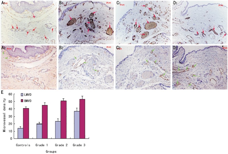
Figure 2. Immunohistochemical staining for blood and lymphatic vessels in petrygia.
There was a number of CD31(+)LYVE-1(−) blood vessels but only few CD31(+)LYVE-1(+) lymphatic vessels in controls. Lymphatic vessels were increased gradually in Grade 1 and 2 pterygia but were dramatically increased in Grade 3 pterygia. (A: controls; B: Grade 1 pterygia; C: Grade 2 pterygium; D: Grade 3 pterygia.;upper panel correspond to CD31 and lower panels to LYVE-1 staining; E: BMVD and LMVD in each group; Red arrows: blood vessels; Green arrows: lymphatic vessels. Magnification for immunohistochemistry ×200).

