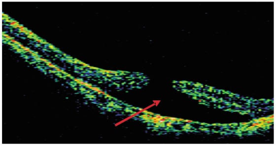
Figure 2. OCT image of a high myopia macular hole accompanied by shallow retinal detachment.

(Patient 3, female, 46 years old, right eye). The solid arrow shows shallow retinal detachment, and the low echo cavum is smooth.

(Patient 3, female, 46 years old, right eye). The solid arrow shows shallow retinal detachment, and the low echo cavum is smooth.