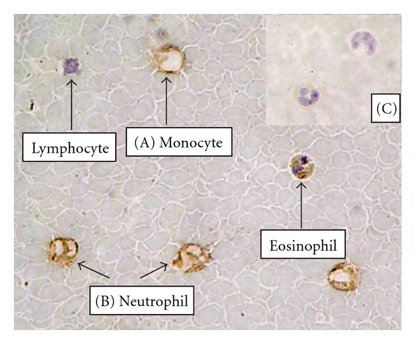
Figure 1.

Immunohistochemical analysis of visfatin protein in WBC cells showed positive brown staining evident of monocyte (A) and neutrophil (B) (magnification, ×1000). Staining was absent in the control section, in which the primary antibody was replaced with nonimmune antiserum (C; magnification, ×400).
