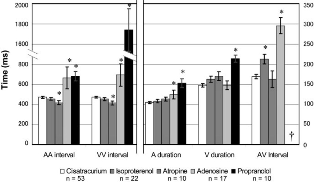
Fig. 5.
Cardiac intervals in stage 46 X. laevis embryos treated with chronotropic medications as measured by video analysis. *Statistically significant difference (P < 0.05 by Tukey’s F test) compared with the cisatracurium paralyzed control group (n = 53; open bars). Left, time scale AA interval and VV interval. Right, time scale for the A duration, V duration, and AV interval. †No measurable AV interval due to second- or third-degree heart block.

