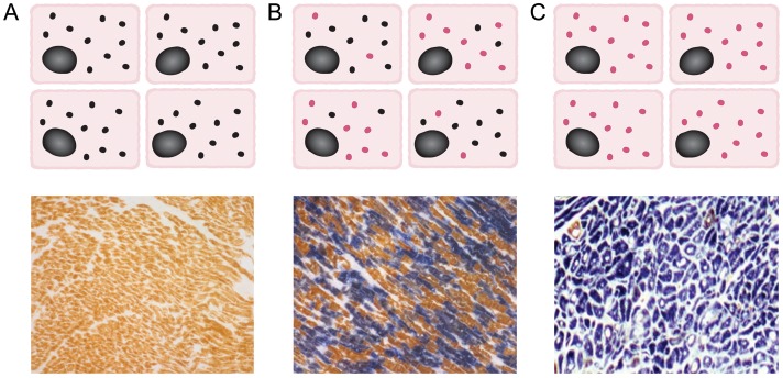
Figure 2.
Mitochondrial DNA mutations and patterns of cellular respiratory function. (A) In normal individuals, all cardiomyocytes contain multiple copies of wild-type mitochondrial DNA (black circles, upper panel), with sequential cytochrome c oxidase/succinate dehydrogenase histochemistry showing all cardiomyocytes as cytochrome c oxidase-positive (brown, lower panel). (B) In patients with heteroplasmic mitochondrial DNA mutations, different proportions of wild-type (black) and mutated mitochondrial DNA (red) are present in individual cardiomyocytes (upper panel); cytochrome c oxidase/succinate dehydrogenase histochemistry reveals a mosaic pattern of cytochrome c oxidase-deficient and cytochrome c oxidase-positive cardiomyocytes, with cellular respiratory deficiency only apparent when a threshold proportion of mutated mitochondrial DNA is reached (lower panel). (C) In patients with homoplasmic mitochondrial DNA mutations, all cardiomyocytes contain multiple copies of mutated mitochondrial DNA (red, upper panel), with the majority of cells displaying cytochrome c oxidase deficiency (blue, lower panel).

