


Figure 2.
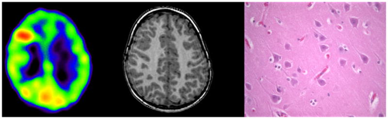
AMT PET scan (left) showing increased tracer uptake in right frontal lobe in a 3 years old boy with intractable seizures and normal MRI (middle). Post-surgical histopathology (right: 40x, H&E) was also normal. The child is having persistent seizures after the surgery.