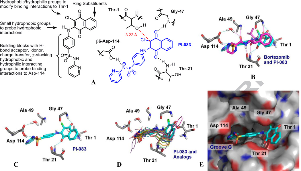
Figure 2.
A. Modifications around PI-083 for library synthesis and predicted binding interactions of PI-083 in the β5 and β6 subunits of the 20S proteasome. B. Overlay of PI-083 (cyan, docked pose) with the Bortezomib (magenta, X-ray crystal pose) in the β5 and β6 units of the 20S proteasome C. PI-083 overlaid in the β5 and β6 subunits of the 20S proteasome. D. PI-083 (cyan, stick representation) and analogs (line representations) shown in Table 1 overlaid in the β5 and β6 subunits of the proteasome. E. Surface model of the 20S proteasome with PI-083. The protein surface of the proteasome is colored according to electrostatic potential; positively charged areas are colored in blue, and negatively charged areas are colored in red. For PI-083, carbon atoms are colored in cyan, oxygen in red, nitrogen in blue, hydrogen in white, and sulfur in yellow. Images were created by PyMol20.

