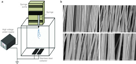
Figure 3.
Electrospinning instrumentation and SEM images of PCL nanofiber scaffolds. (a) The instrumentation consists of a syringe with spinneret, a syringe pump, a high voltage power supply, and a nanofiber gap collector. (b) Scanning electron microscopy (SEM) images of paralleled aligned PCL nanofiber scaffolds that were taken using a field-emission scanning electron microscope (S4700; Hitachi, Tokyo, Japan). Nanofibers were electrospun on the gap collector by adjusting flow rate of PCL solution at 0.5 ml/hour using a syringe pump while a potential of 12 kV was applied between the spinneret (a 22-gauge needle) and a grounded collector located 12 cm away. NT, nontranscribed; PCL, polycaprolactone.

