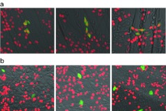
Figure 7.
DNA replication activity of eGFP+ and eGFP− cells plated onto polylysine-coated nanofiber scaffolds or polylysine-coated dish surfaces. (a) HCT116-19 cells (1 × 10⁶) that were targeted with 72 NT for gene editing were plated onto a polylysine-coated PCL nanofiber scaffold for 72 hours. DNA replication activity was measured using the Click-iT EdU assay. The genetically modified cells (eGFP+) exhibit green fluorescence, and cells with active DNA replication display red due to incorporation of EdU into newly synthesized DNA. (b) HCT116-19 cells that have undergone genetic modification as described above were plated onto a polylysine-coated 6-well dish surface. The Click-iT EdU assay was carried out 72 hours after plating to measure the DNA replication activity, and the images were taken using EVOS FL microscope (AMG Micro). eGFP, enhanced green fluorescent protein; NT, nontranscribed; PCL, polycaprolactone.

