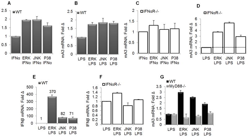
Figure 8. MAPK family members are negative regulators of mA3 in primary macrophages.
(A, B, and E) primary IFNαR WT BMDM (C, D, and F) primary IFNαR−/− BMDM on C57BL/6 background were left untreated or pre-treated with inhibitors of ERK (10μM), JNK (90 nM), and P38 (50 nM) for 15 minutes at 37°C followed by stimulation with 1000 units/ml of endotoxin-free recombinant IFNα for 4 hours, 1μg/ml LPS for 6 hours, or vehicle. Cells were used for RNA extraction, cDNA synthesis and qPCR evaluation of (A, B, C, and D) mA3 mRNA levels or (E and F) IFNβ mRNA. (G ) Immortalized wild-type and MyD88−/− BMDM on C57BL/6 background were left untreated or pre-treated with inhibitors of ERK (10μM), JNK (90 nM), and P38 (50 nM) for 15 minutes at 37°C followed by stimulation with 1000 units/ml of endotoxin-free recombinant IFNα for 4 hours, 1μg/ml LPS for 6 hours, or vehicle. Cells were used for RNA extraction, cDNA synthesis and qPCR examination of mA3 mRNA levels. Data were normalized to GAPDH and presented as fold change relative to vehicle treated cells. Error bars are standard error, * is significance with p value less than 0.05 and ** is significance with p value less than 0.01. The numbers on figure E denote mRNA values. Experiments were repeated at least three different times with similar results.

