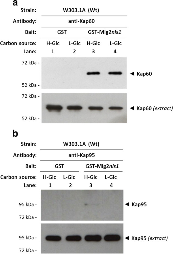
Figure 5.
Interaction of Kap60 and Kap95 with Mig2 nls1. The GST-Mig2nls1fusion protein was purified on glutathione-Sepharose columns. Equal amounts of GST-Mig2nls1 were incubated with cell extracts from the wild-type strain W303-1A. The yeasts were grown in YEPD media until an A600nm of 0.8 was reached and then shifted to low (L-Glc) glucose conditions for 1 h. After exhaustive washing the proteins were separated by 12% SDS-PAGE, and retained Kap60 and Kap95 proteins were visualized on a Western blot with polyclonal anti-Kap60 (b) and anti-Kap95 (c) antibodies respectively. For the control samples, GST protein was also incubated with high- (H-Glc) and low-glucose (L-Glc) cell extracts, but no signals were detected. The level of Kap60 and Kap95 proteins present in the different extracts used in Figure 5a and 5b was determined by Western blot using anti-Kap60 and anti-Kap95 antibodies respectively. The Western blots shown are representative of results obtained from four independent experiments.

