Figure 1.
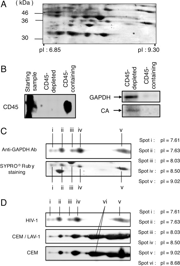
Confirmation of GAPDH incorporation into HIV-1 virions. (A) SYPRO® Ruby-stained 2D gel image of proteins from highly purified HIV-1LAV-1 virions within the pI range of 6.85-9.30 and the molecular weight range of 30–46 kDa. (B) Western immunoblotting of GAPDH in CD45-depleted fraction and CD45-containing fraction. CD45 and CA levels were used as the markers of microvesicles and viral particles, respectively. Samples from the CD45-immunoaffinity-depleted fraction (CD45-depleted fraction) and anti-CD45 beads fraction (CD45-containing fraction) are identified above their respective lanes. The antibodies used are indicated on the left side of each blot. (C) Detection of virion-associated GAPDH isozymes. The upper panel shows the results of western immunoblotting using an anti-GAPDH antibody and the lower panel indicates the corresponding SYPRO® Ruby-stained gel. (D) Comparison of pI values of GAPDH isozymes among HIV-1LAV-1 (upper panel), CEM/LAV-1 (middle panel), and CEM cells (lower panel). GAPDH isozymes were detected by western immunoblotting using the anti-GAPDH antibody. Spot pI values are shown in the figure.

