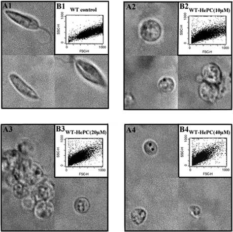
FIG. 2.
Morphology and cell size of L. donovani WT promastigotes cultured for 24 h in fresh complete medium at 26°C in the absence or presence of different concentrations of HePC. The results of light microscopy (magnification, ×1,000) (A) and flow cytometry analysis (B) of the cell sizes of WT promastigotes either untreated (A1 and B1) or exposed to 10 μM (A2 and B2), 20 μM (A3 and B3), or 40 μM HePC (A4 and B4) are shown.

