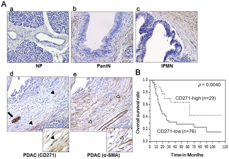
Figure 1. Characterization of stromal CD271 expression in pancreatic cancer.
(A) Immunohistochemical staining for CD271 in pancreatic tissues. (A-a) In normal pancreas (NP), stromal cells rarely stain positive for CD271. (A-b, -c) Stromal cells are strongly stained around pancreatic intraepithelial neoplasia (PanIN) (b) and intraductal papillary mucinous neoplasm (IPMN) (c). (A-d) In pancreatic ductal adenocarcinomas (PDAC), stromal cells are partly stained, and CD271+ stromal cells are not adjacent to pancreatic cancer cells. Black arrowheads and arrows indicate CD271+ cells and nerves as positive control in the serial sections. (A-e) α-smooth muscle actin (SMA) is expressed most stromal cells around cancer cells and neoplastic tubules. White arrowheads indicate α-SMA cells in the serial sections. Original magnification: 200×, insets: 600×. (B) Kaplan–Meier survival analysis for CD271 high expression in the stroma of PDAC. Stromal CD271 high expression is associated with a good prognosis (p = 0.0040).

