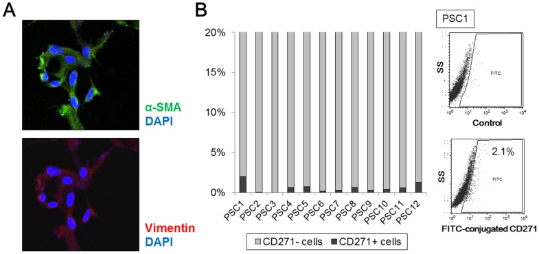
Figure 2. Analyses of CD271 expression in human pancreatic stellate cells (PSCs) isolated from pancreatic tissues.
(A) Representative microphotograph of immunofluorescence staining for α-smooth muscle actin (SMA) (green) and vimentin (red) in PSCs. Nuclei were counterstained with 4′,6-diamidino-2-phenylindole (blue). PSCs show a stellate-like or spindle-shaped morphology and express α-SMA. Original magnification: 200×. (B) The positive rates of CD271 expression in PSCs are 0.0–2.1% by flow cytometric analyses. Representative flow cytometric images of CD271 in activated PSCs are also shown (right).

