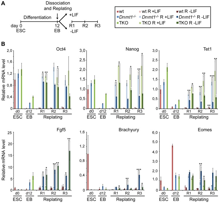
Figure 6. LIF signaling induces reversion of cells from Dnmt1−/− and TKO EBs to the ESC state.
(A) Overview of experimental set up. Day 12 EBs were dissociated and their cells were plated and further cultured for three days (R1–3) in the presence or absence of LIF. (B) Transcript levels of pluripotency associated genes Oct4, Nanog and Tet1, as well as differentiation marker genes Fgf5, Brachyury and Eomes were determined by RT-qPCR in undifferentiated ESCs (day 0), day 12 EBs and 1, 2 and 3 days after replating (R1–3). Wild type, Dnmt1−/− and TKO samples are represented in shades of red, blue and green, respectively, as indicated in the box at the upper right corner. Mean values relative to wt ESCs (day 0) and standard errors are from three independent biological replicates. Asterisks indicate significance levels: * p<0.05; ** p<0.001; *** p<0.0001 (Student t-test).

