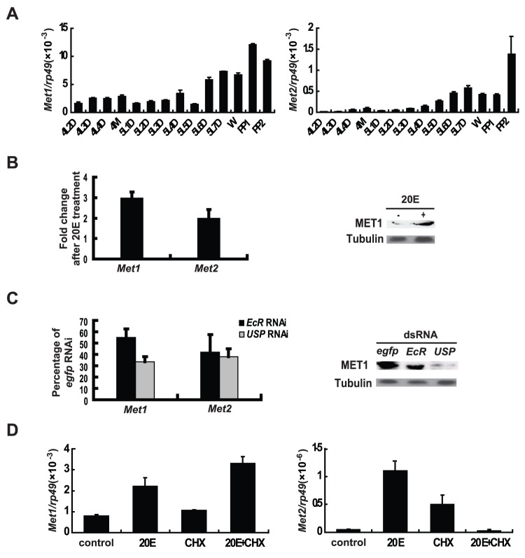
Figure 1. Two Met genes in the Bombyx genome.
Three biological replicates were used, one of which is represented. In each biological replicate, more than 10 larvae were used (A–C). (A) From day 2 of the 4th instar to day 2 of the prepupal stage, Met1 and Met2 mRNA expression in fat body was determined by qPCR. The developmental profiles show expression peaks during molting and pupation. 4L2D, day 2 of the fourth instar, and so on; M, molting; W, the wandering stage; PP, the prepupal stage. (B) Met1 and Met2 mRNA levels (left panel), and MET1 protein level (right panel) were increased by 20E treatment in vivo. 20E (1 µg per larva) was injected into selected larvae on day 2 of the fifth instar, and fat body was explanted for qPCR analysis and Western blots 6 hr after 20E treatment. Tubulin was used as a loading control. (C) Met1 and Met2 mRNA levels (left panel), and MET1 protein level (right panel) were decreased by EcR RNAi and USP RNAi in vivo. dsRNA (10 µg per larva) was injected into larvae during the initiation of the early wandering stage, and fat body was explanted for qPCR analysis and Western blots 24 hr after RNAi treatment. Tubulin was used as a loading control. (D) Simultaneous addition of 1 µM 20E and 10 µg/ml cycloheximide (CHX) to Bombyx DZNU-Bm-12 cells for 2 hr revealed that Met1 and Met2 are 20E primary- and secondary-response genes, respectively.

