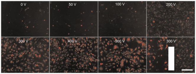
Figure 1. Efficiency of electroporation in HMEC-1 monolayers.
Pictures of monolayers were taken within 3 min post-EP in electroporation buffer containing 100 µM PI. The vertical bar indicates the position of the right parallel electrode: area to the left within the E of 300 V and area to the right outside the electric field. Bar = 200 µm.

