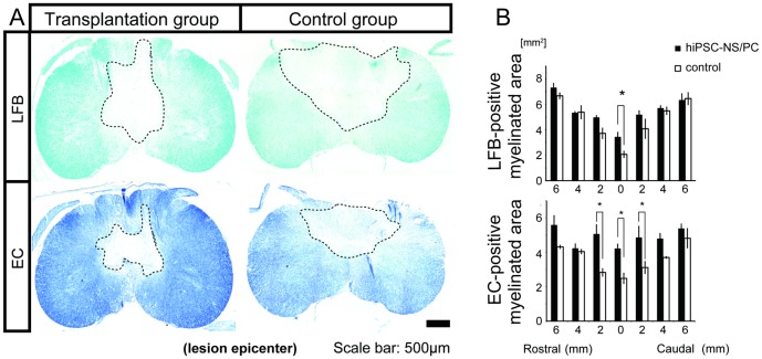
Figure 2. Grafted hiPSC-NS/PCs prevent demyelination after SCI.
(A) Representative LFB- and EC-stained images of axial sections through the lesion epicenter at 12 weeks post-engraftment. The region enclosed by the dashed line indicates the demylinated area. (B) Quantification of the images revealed significant preservation in the size of the myelinated areas at the lesion epicenter in the transplantation group compared with the vehicle control group. Data represent the mean ± SEM (n = 3 per group, *p<0.05).

