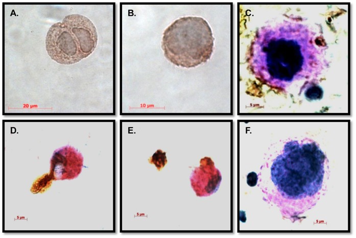
Figure 3. Megakaryocytes were likely the dominant dengue virus antigen positive cells in monkey bone marrow.
Smears of bone marrow cells were prepared and immunohistochemical stainings were performed as described in Methods. Dengue antigen (4G2 positivity) is indicated by DAB staining (brown) (A) Dengue viral antigen in a diploid megakaryocyte. (B) Dengue antigen in a multi-lobulated megakaryocyte. (C), Dengue antigen in cellular debris. Red, PAS staining. Blue, hematoxylin staining. (D and E) Dengue viral antigen-containing vesicles engulfed by phagocytic cells. (F) Isotype control.

