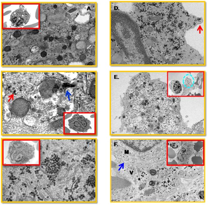
Figure 8. Viral particles are present in megakaryocytes from the human bone marrow.
Sample preparations for EM studies were performed as described in the Methods. (A) Uninfected control. (B) Cellular vesicle containing viral particles (single particle, red arrow; cluster of viral particles, blue arrow) inside a diploid megakaryocyte on day one post-infection. (C) Large numbers of viral particles inside the cytoplasm of a multi-lobulated megakaryocyte on day three post-infection. (D) Cytoplasm containing many virus particles shedding off in a vesicle (red arrow). (E) A virion-containing vesicle (dash circle) at the vicinity of an activated mononuclear cell. (F) Virion containing vesicle (V) fusing with a monocyte (M). A zipper junction (blue arrow) is indicated. No viral particles were observed in the monocytes. A scale bar is 0.2 µM.

