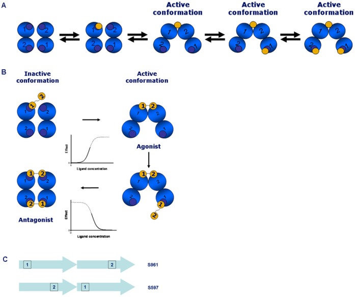
Figure 6. Current model of IR activation and proposed binding mechanism for S961. A.
. Current model of IR activation. The four blue circles represent the receptor binding sites (sites 1 and 2) seen from a top view. Insulin is depicted as a yellow circle. For a detailed explanation of binding sites 1 and 2, see [24]. B. Proposed binding mechanism for S961. The four blue circles represent the receptor binding sites (sites 1 and 2) seen from a top view. For a detailed explanation of binding sites 1 and 2, see [24]. The S961 peptide (Site 1–2 peptide) is shown as two connected yellow circles. At concentrations of 1–10 nM, S961 crosslinks the receptor, leading to agonist activity. At concentrations of above 10 nM, the higher flexibility of S961 in comparison to the insulin molecule allows simultaneous crosslinking of both pairs of binding sites, leading to an inactive conformation and antagonism. The corresponding activation and inactivation sigmoids are also shown. C. Orientation of peptide binding sites. If site 1 is located N-terminally and site 2 C-terminally, a longer distance between the binding sites in S961 in comparison to S661 can be achieved.

