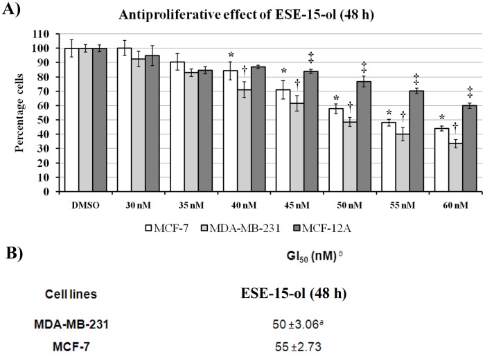
Figure 3. Antiproliferative effects of ESE-15-ol on MDA-MB-231, MCF-7 and MCF-12A cells after 48 h exposure.
A) Dose-dependent analysis of DNA content revealed a statistically significant 50% growth inhibitory effect (GI50) of malignant cell numbers after 48 h of exposure to be 55 nM of ESE-15-ol for in MCF-7 and 50 nM for the MDA-MB231. MCF-12A cells were the least affected at 50 nM of ESE-15-ol. a Values are presented as the average of three biological replicates (N = 6) ± SD. b The compound concentration required to inhibit cell proliferation by 50% was determined by exposing cells for 48 hours to test inhibitors. * indicates a t-test P-value <0.05 for growth inhibition between MCF-7 and MDA-MB-231 cells. † indicates a t-test P-value <0.05 for growth inhibition between MDA-MB-231 and MCF-12A cells. ‡ indicates a t-test P-value <0.05 for growth inhibition between MCF-12A and MCF-7 cells.

