
Figure 2. Slug, Twist and Zeb1 localization during mammary gland morphogenesis.
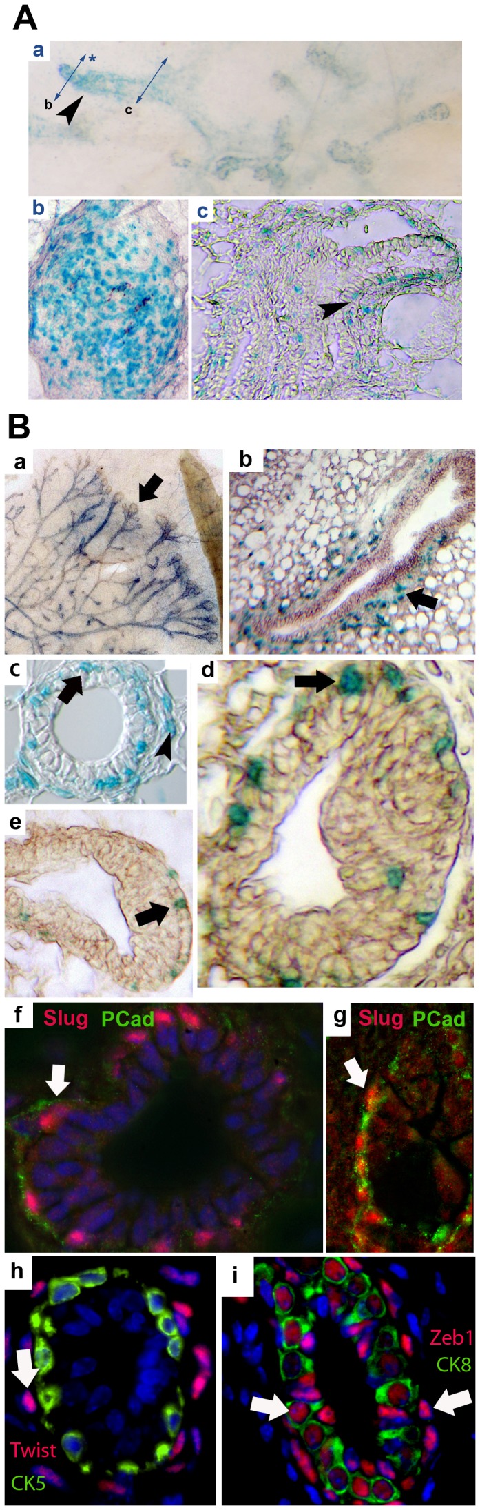
A. At 3 weeks, Slug expression (blue X-Gal staining) is visible in the primary duct on a mammary gland wholemount (a, arrowhead), and section (b). Lower sectioning level (c) show Slug expression in polarizing basal epithelial cells during initial tubulogenesis (arrowhead). B. During mammary tubulogenesis at 6 weeks, Slug is found in growing tubules in wholemount (a) and tubule sections (b–c) involving epithelial cells (c, arrow) and peri-tubular mesenchymal cells (b, arrow and c, arrowhead). Cell location is better defined at higher magnification, including basal epithelial (arrow) and mesenchymal (arrowhead) cells (c). Tubule terminal end buds (TEB) sections also show expression by mostly basal epithelial cap cells (d–e, arrows). Co-labeling using DAPI (blue) and antibodies against Slug (red) and Pcad (P-cad, green) demonstrates a basal localization for Slug in 6 weeks (f) and adult (g) tubules. In addition, Twist (h) and Zeb1 (i) were located in tubules from 10 weeks-old mammary glands. Colabeling with CK5 show that only stromal cells express Twist (arrow). Conversely, CK8 colabeling show that Zeb1 is expressed by basal and luminal epithelial cells (arrows).