
Figure 5. Slug controls mammosphere growth.
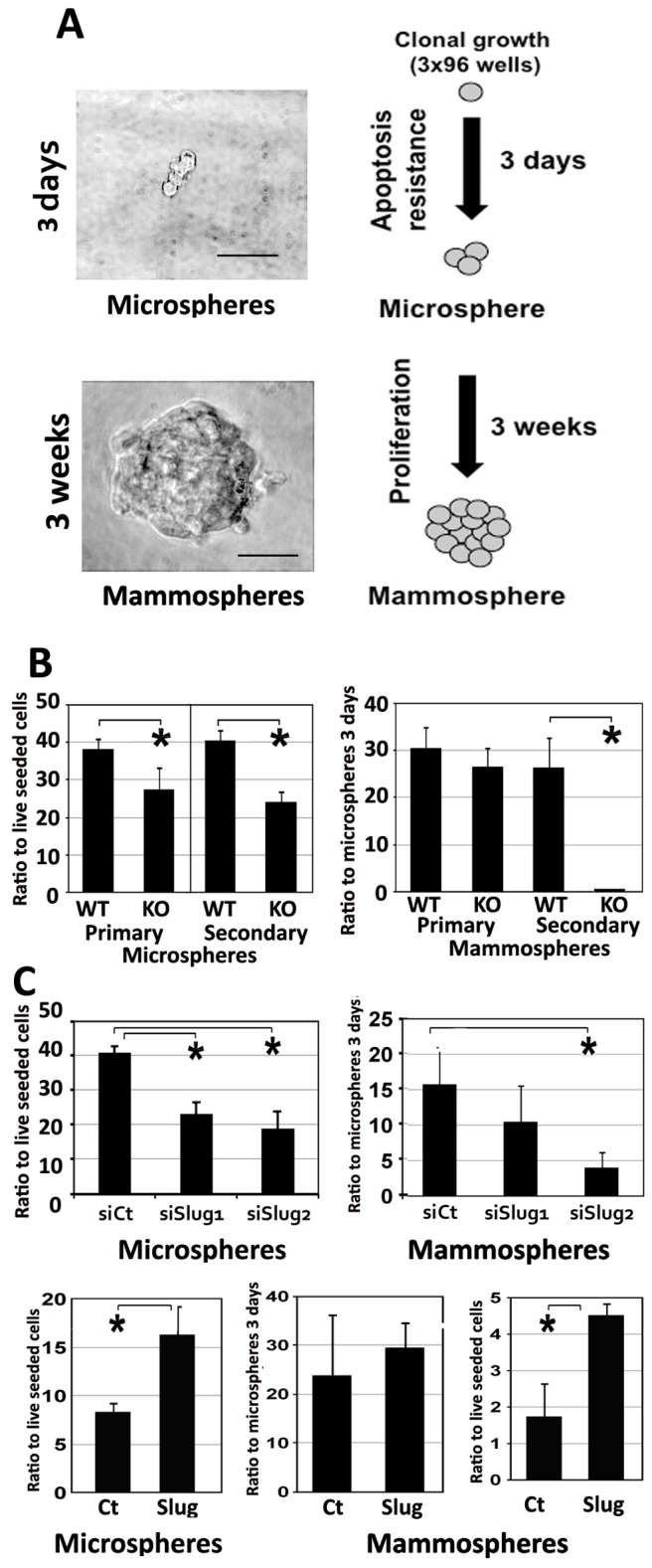
A. Epithelial cells isolated from mammary glands were seeded individually in low-adherence culture wells. All wells were screened after 3 days. Wells containing 1–4 live cells (microsphere) were enumerated. After three weeks, Some of the microspheres evolved into mammospheres that were monitored. Values are reported as percentage compared to the number of live seeded cells or to the number of microspheres at 3 days. Bar = 50 µm. B. Cells from wild-type (WT) or Slug-deficient mouse (KO) were cloned to grow into primary microspheres and mammospheres. For secondary mammospheres, 10–20 primary mammospheres were pooled and enzymatically dissociated before individual cloning and further screening. Experiment was repeated three times. C. CommaDβ cells were transfected with control (siCt) or two distinct anti-Slug siRNA (siSlug1 and siSlug2) and seeded individually in 96 well low-adherence dishes. All wells were screened after 3 days. Microspheres and mammospheres were reported separately after 3 weeks. A significant decrease in viability was observed after 3 days in cells transfected with anti-Slug siRNA. This deficit was even stronger three weeks later (*Student test, p<0.05). CommaDβ cells were also transfected with control (Ct) or Slug full-length cDNA (Slug) expression vectors and seeded individually in 96 well dishes as previously. The number of mammospheres showed a significant and early increase in survival and growth for Slug-overexpressing mammospheres (*Student test, p<0.05).